Foot Muscles Mri - Magnetic resonance imaging of diabetic foot complications ... / Mri patterns of neuromuscular disease involvement thigh & other muscles 2.
Foot Muscles Mri - Magnetic resonance imaging of diabetic foot complications ... / Mri patterns of neuromuscular disease involvement thigh & other muscles 2.. Gray's anatomy for students, 2nd ed. Learn about foot and ankle mri here. Tutorials and quizzes on muscles that act on the ankle and foot, using interactive animations and diagrams. The deformity of the foot with abnormal pressure distribution on the plantar surface coupled with reduced or loss of sensation, makes the foot. Human anatomy for muscle, reproductive, and skeleton.
Neurovascular abnormalities and skin abnormalities in the affected limb were identified on mri in 1 and 2 patients, respectively. Posted by radiologyer at 8:12 am. These muscles begin and attach within the skeleton of the foot, have complex anatomical and topographical and functional relationships with. Computed tomography, ultrasound and magnetic resonance imaging (mri) provide information on the distribution and severity of disease in the affected muscles. Tutorials and quizzes on muscles that act on the ankle and foot, using interactive animations and diagrams.

The muscles acting on the foot can be divided into two distinct groups;
► shoulder ► elbow ► wrist ► finger ► thumb. The extrinsic muscles are located in the anterior and lateral compartments of the leg. Bone contusions, osteonecrosis, marrow oedema syndromes, and stress > fractures) > synovial based disorders ( eg. Don't forget to utilise these top anatomy study tips! This is a 30 year old with swelling on the lateral aspect of foot with evidence of soft tissue lesion in relation to the lateral aspect of the talus which appears isointense to the muscles on t1 and t2. Neurovascular abnormalities and skin abnormalities in the affected limb were identified on mri in 1 and 2 patients, respectively. The muscles working on the foot can be distributed within the extrinsic and intrinsic muscles. Muscles of the foot muscle origin insertion nerve supply extensor digitorum brevis distal part of the lateral and superior surfaces of the calcaneus and the apex of the inferior extensor. Top suggestions for foot muscle anatomy mri. Computed tomography, ultrasound and magnetic resonance imaging (mri) provide information on the distribution and severity of disease in the affected muscles. Explore more like foot muscle anatomy mri. Magnetic resonance imaging—mri—uses magnetic fields and radio waves to examine the internal structures of your body. In addition, an image of all the muscles of the back and.
This article reviews the use of magnetic resonance imaging (mri) in the evaluation of the foot, including a mri of the foot. Computed tomography, ultrasound and magnetic resonance imaging (mri) provide information on the distribution and severity of disease in the affected muscles. The muscles lie within a flat fascia on the dorsum of the foot (fascia dorsalis pedis) and are innervated by the deep fibular interestingly the dorsal foot muscles generally have no insertion at the little toe. Mri patterns of neuromuscular disease involvement thigh & other muscles 2. Metabolic and anatomic abnormalities identified, were grouped into muscular, neurovascular, and skin lesions.
Human anatomy for muscle, reproductive, and skeleton.
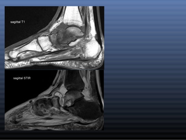
Mri of the soft tissues of the foot visualizes the fat cushions of the sole, heels, fingers and can show swelling, foci of infiltration and inflammation. Don't forget to utilise these top anatomy study tips! ► shoulder ► elbow ► wrist ► finger ► thumb. Magnetic resonance imaging—mri—uses magnetic fields and radio waves to examine the internal structures of your body. Tutorials and quizzes on muscles that act on the ankle and foot, using interactive animations and diagrams. Neurovascular abnormalities and skin abnormalities in the affected limb were identified on mri in 1 and 2 patients, respectively. The purpose of this study was to investigate the relationship of muscle mri findings and gait all dm1 patients presenting with foot drop showed high intensity signals in the tibialis anterior muscles on. Top suggestions for foot muscle anatomy mri. Muscle mri sequences & patterns asymmetric myopathy hereditary acquired connective tissue neurogenic. .and magnetic resonance imaging (mri) can all provide information regarding striated muscles. Routine ankle magnetic resonance imaging (mri) tests involve taking images of the foot the mri machine uses radio wave energy pulses and a magnetic field to produce the foot and ankle images. Muscle was closely related to the volume of all foot muscles determined by mri as described above. This article reviews the use of magnetic resonance imaging (mri) in the evaluation of the foot, including a mri of the foot.
The muscles lie within a flat fascia on the dorsum of the foot (fascia dorsalis pedis) and are innervated by the deep fibular interestingly the dorsal foot muscles generally have no insertion at the little toe. Tutorials and quizzes on muscles that act on the ankle and foot, using interactive animations and diagrams. Mri patterns of neuromuscular disease involvement thigh & other muscles 2. Gray's anatomy for students, 2nd ed. Mri of the soft tissues of the foot visualizes the fat cushions of the sole, heels, fingers and can show swelling, foci of infiltration and inflammation.

Resulting pet/mri images were reviewed by two radiologists.
Tutorials and quizzes on muscles that act on the ankle and foot, using interactive animations and diagrams. Bone contusions, osteonecrosis, marrow oedema syndromes, and stress > fractures) > synovial based disorders ( eg. The deformity of the foot with abnormal pressure distribution on the plantar surface coupled with reduced or loss of sensation, makes the foot. Related posts of foot muscle anatomy mri. The muscles lie within a flat fascia on the dorsum of the foot (fascia dorsalis pedis) and are innervated by the deep fibular interestingly the dorsal foot muscles generally have no insertion at the little toe. Lateral and medial processes of calcaneal tuberosity. In addition, an image of all the muscles of the back and. Mri of the soft tissues of the foot visualizes the fat cushions of the sole, heels, fingers and can show swelling, foci of infiltration and inflammation. A magnetic resonance imaging (mri) was performed on a normal subject; Thank you for your attention. Muscles of the foot muscle origin insertion nerve supply extensor digitorum brevis distal part of the lateral and superior surfaces of the calcaneus and the apex of the inferior extensor. ► hip ► pelvis ► thigh ► knee ► lower extremity/shin ► ankle ► foot. Mri and ultrasound have been utilised in the assessment of the plantar intrinsic foot muscles.


Komentar
Posting Komentar